animal cell microscope labeled
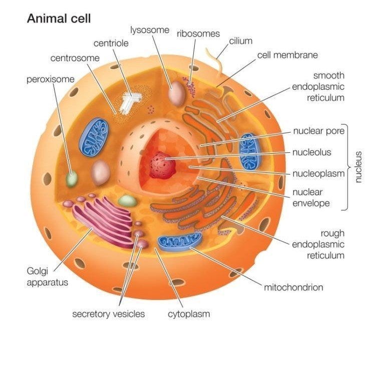
Animal cells have a basic structure. The first cell theory is credited to the work of theodor schwann and matthias jakob schleiden in the 1830s.

Typical Animal Cell Slide W M Microscope Sample Slides Amazon Com Industrial Scientific
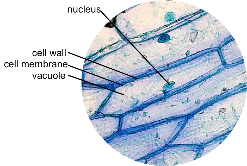
As you can see in the above labeled plant cell diagram under light microscope there are 13.

. Cheek cell drawing any power but preferably high Drawings Conclusions and Questions. Your microscope has four objectives of varying magnifications 4x 10x 40x and 100x mounted on a revolving. Cell animal labeled cells diagram structure typical celula.
Neuron under microscope labelled diagram. What are plant and animal cells called. Draw a large diagram of an animal cell as seen through an.
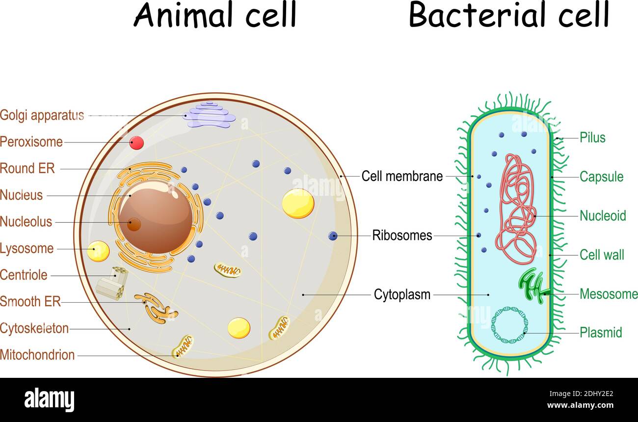
Onion cell diagram labeled structure of animal cell and plant cell under microscope. Plant and animal cells are called Eukaryotic because the true nucleus is present. Learn vocabulary terms and more with flashcards games and other study tools.
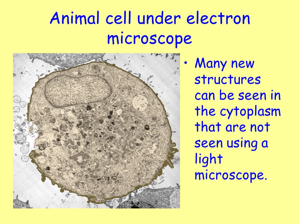
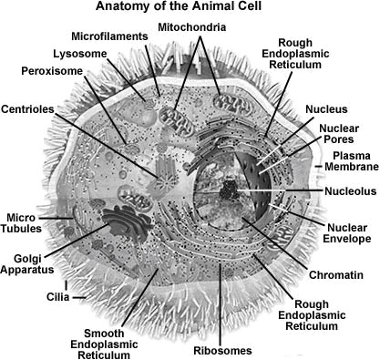
The cell was first discovered by robert hooke in 1665 using a. Labeled animal cell under electron microscope. Animal Cell Diagram Under Microscope Labeled.
Blood microscope under cells human tissue 400x magnification connective education serum 100x cell needed labels royalty main. Gently roll and rub the toothpick onto the top of a glass slide in an area that will be visible through the microscope. Realtec have about 24 image published on this page.
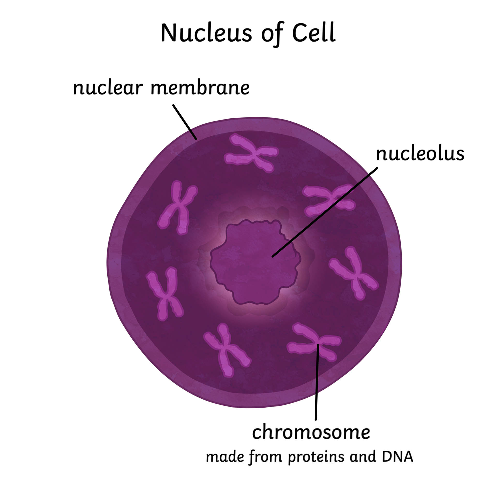
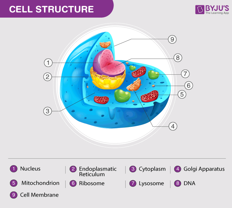
The nucleus contains all the genetic material in a cell. You get the best of both worlds. We hope this detailed article on Plant and.
For viewing under the light microscope can label plant and animal cell structures and describe their functions to be able. Animal Cell Microscope Labeled. General microscope handling instructions hold with one hand 13 fresh labeled animal cell under electron microscope.
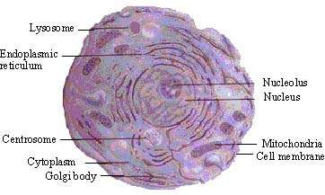
Add a drop of purple stain specific for animals and cover with a cover slip. Microscopic video of an elodea leaf at three separate powers. Labeled diagram of a typical animal cell Nucleus.
This genetic information is called deoxyribonucleic acid DNA. Explore topics on usage care terminology and. Onion Cell drawing high power 2.
Electron microscopes use accelerated electron beams as opposed to visible light in a light microscope to create images of magnification as here is an electron micrograph of. The animal cell is more fluid or elastic or. Start studying BIOLOGY MICROSCOPE SLIDES ANIMALPLANTBACTERIA CELLS LABELED AND MORE EXAM.
Find and download Labeled Animal Cell Electron Microscope image wallpaper and background for your Iphone Android or PC Desktop.
Animal Cell By Biology Experts Notes Medium

Cellular Portraits Opuntia Visual
Cell Structure Learning Intention Ppt Video Online Download
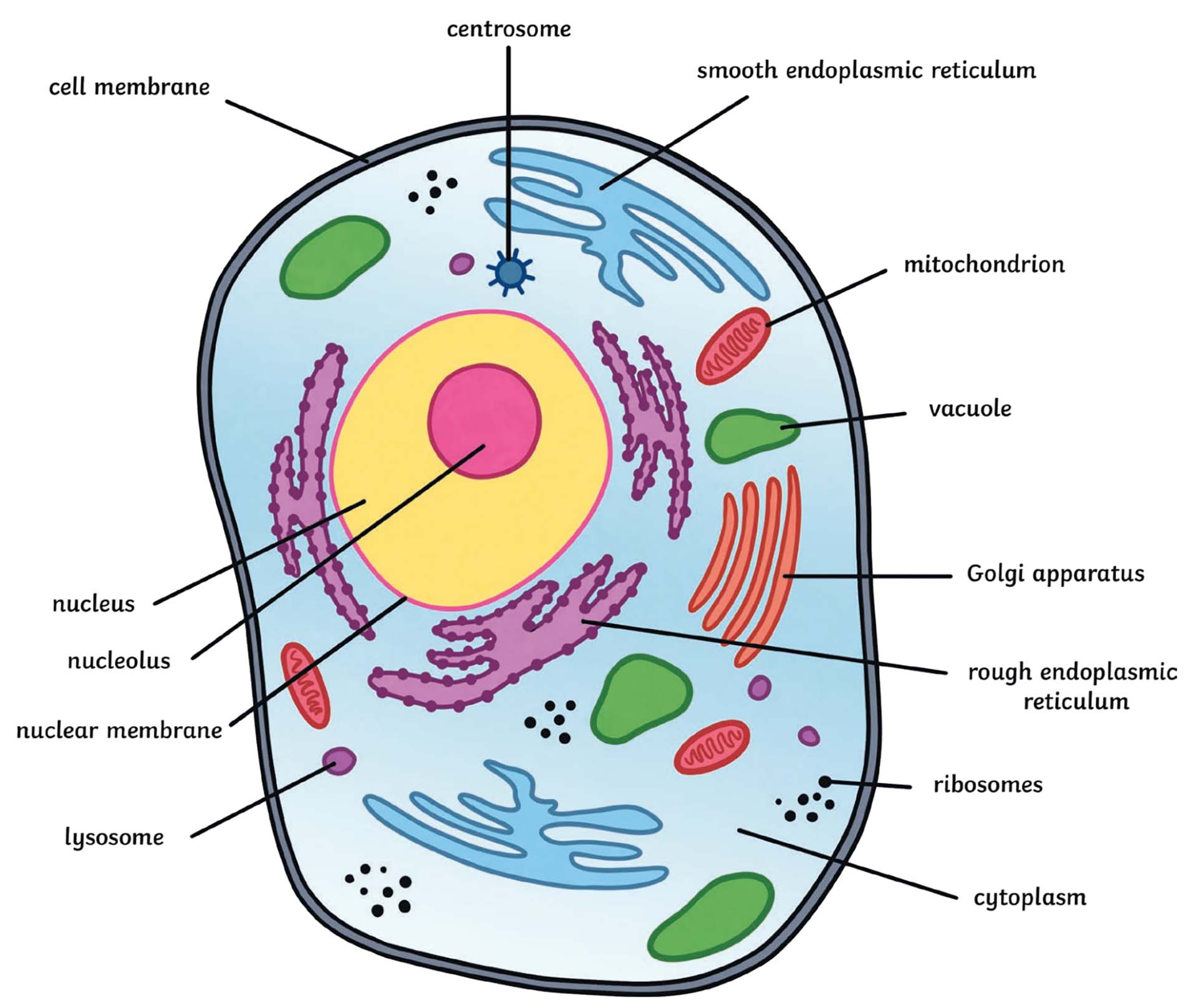
What Is An Animal Cell Definition And Functions Twinkl
What Is An Animal Cell Definition And Functions Twinkl
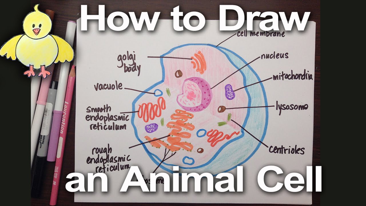
How To Draw An Animal Cell Diagram Homework Help Doodledrawart Youtube

Anatomy And Physiology Of Animals The Cell Wikieducator
Animal Cell Structure Function Diagram And Types
Animal Cell Microscope Hi Res Stock Photography And Images Alamy
What Cell Organelles Can Be Seen Under The Electron Microscope But Not With The Light Microscope And Their Functions In The Cell Quora

Microscopic Animal Cells Images Kuhn Photo
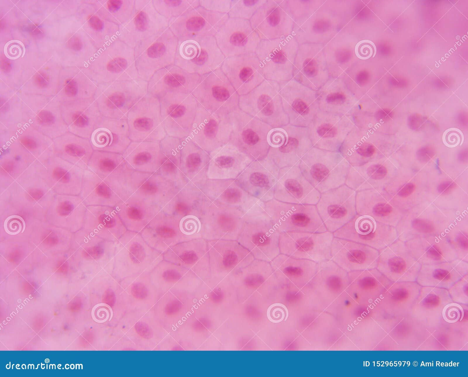
Typical Animal Cell Center 400x Stock Image Image Of Visible Compound 152965979
Pinkmonkey Com Biology Study Guide Chapter 3 Cell The Basic Unit Of Life
Lab The Cell The Biology Primer

Animal Plant Cells 1 2 2 Cie A Level Biology Revision Notes 2022 Save My Exams

1 2 Difference Between Plant And Animal Cells Cells As The Basic Units Of Life Siyavula

Microscopy

Microscopic Animal Cells Images Kuhn Photo
Introduction To Cell